What is a Light Microscope?
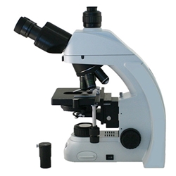
A light microscope is a tool that enlarges and allows for the observation of tiny objects, often those that are not visible to the naked eye. It employs visible light and a series of lenses to achieve magnification and resolution. Here are the key components and features of a light microscope:
1. Illumination Source: A light source, usually a built-in lamp, provides the necessary light to illuminate the specimen. In some cases, natural light or an external light source can also be used.
2. Condenser Lens: This lens focuses the light onto the specimen, enhancing the illumination and contrast.
3. Objective Lenses: Located near the specimen, these lenses magnify the image of the specimen. Most light microscopes have multiple objective lenses with different magnification powers (e.g., 4x, 10x, 40x, 100x).
4. Ocular Lenses (Eyepieces): These lenses are located at the top of the microscope and further magnify the image formed by the objective lenses. They typically have a magnification power of 10x.
5. Stage: The platform where the specimen slide is placed. The stage can be adjusted to move the slide horizontally and vertically for proper positioning under the objective lens.
6. Focus Knobs: Coarse and fine adjustment knobs are used to bring the specimen into sharp focus. The coarse focus knob moves the stage or objective lenses quickly, while the fine focus knob allows for precise focusing.
7. Diaphragm or Iris: This component controls the amount of light reaching the specimen, which can be adjusted to improve contrast and resolution.
Light microscopes are widely used in various scientific fields, including biology, medicine, and materials science. They allow researchers to observe the structure, morphology, and behavior of cells, tissues, and microorganisms, as well as to conduct detailed analyses of small-scale structures.
10 Types of Light Microscopes and How They're Used
Light microscopes are essential tools in scientific research, enabling the observation and analysis of microscopic structures. Here, we explore ten types of light microscopes, highlighting their unique features and providing guidance on their use.
1. Stereo Microscopes (Dissecting Microscope)
Overview: Stereo microscopes, also known as dissecting microscopes, provide a three-dimensional view of specimens. They are ideal for examining the surface details of solid specimens at relatively low magnification.
How to Use:
- Place the specimen on the stage.
- Adjust the focus using the coarse and fine adjustment knobs.
- Use the built-in illumination to enhance the visibility of surface features.
- Observe the specimen through the binocular eyepieces.
2. Compound Microscopes
Overview: Compound microscopes use multiple lenses to achieve high magnification, making them suitable for viewing thin sections of specimens. They are widely used in biological and medical research.
How to Use:
- Prepare a thin specimen on a glass slide and cover it with a coverslip.
- Place the slide on the stage and secure it with stage clips.
- Start with the lowest magnification objective lens and use the coarse focus knob to bring the specimen into view.
- Switch to higher magnification lenses and fine-tune the focus as needed.
3. Digital Microscopes
Overview: Digital microscopes are equipped with a digital camera, allowing for the display of images on a computer screen. They can incorporate various imaging techniques, providing versatility in observation and analysis.
How to Use:
- Connect the microscope to a computer using the provided software.
- Place the specimen on the stage and adjust the focus using on-screen controls.
- Capture and save images or videos for further analysis.
4. Bright Field Microscopes
Overview: Bright field microscopes are a standard form of the compound microscope that uses transmitted light to illuminate the specimen, creating a bright background.
How to Use:
- Prepare and place the specimen slide on the stage.
- Use the condenser to focus the light through the specimen.
- Adjust the objective lenses and focus knobs to achieve a clear image.
5. Dark Field Microscopes
Overview: Dark field microscopes enhance contrast by using oblique light, causing the specimen to appear bright against a dark background. This technique is useful for observing unstained specimens.
How to Use:
- Position the specimen slide on the stage.
- Adjust the dark field condenser to direct light obliquely.
- Use the objective lenses and focus knobs to bring the specimen into view.
6. Differential Interference Contrast Microscopes (DIC)
Overview: DIC microscopes use polarized light to enhance contrast in unstained, transparent specimens, providing a pseudo-3D effect.
How to Use:
- Place the specimen on the stage and adjust the DIC prism settings.
- Use the polarizer and analyzer to achieve the desired contrast.
- Focus on the specimen using the objective lenses and adjustment knobs.
7. Polarizing Microscopes
Overview: Polarizing microscopes utilize polarized light to study birefringent materials, commonly used in geology and material science.
How to Use:
- Place the specimen on the stage.
- Adjust the polarizer and analyzer to align with the optical path.
- Rotate the stage to observe changes in the specimen's appearance.
8. Phase Contrast Microscopes
Overview: Phase contrast microscopes enhance contrast in transparent specimens without staining, ideal for viewing live cells.
How to Use:
- Prepare the specimen on a glass slide.
- Align the phase ring in the condenser with the phase plate in the objective lens.
- Adjust the focus to view the specimen with enhanced contrast.
9. Fluorescence/Fluorescent Microscopes
Overview: Fluorescence microscopes use fluorescent dyes to label and study specific components within a specimen, widely used in biological research.
How to Use:
- Stain the specimen with fluorescent dyes.
- Place the specimen on the stage and use the appropriate filter sets.
- Adjust the focus and excitation light to observe the fluorescent signals.
10. Confocal Microscopes
Overview: Confocal microscopes use laser light and fluorescence to produce high-resolution, three-dimensional images, allowing for detailed studies of thick specimens.
How to Use:
- Prepare and stain the specimen as required.
- Use the software to control laser scanning and image acquisition.
- Adjust the focus and settings to capture high-resolution images.
Choosing The Right Microscope for Your Research Needs
Understanding the distinct features and applications of these ten types of light microscopes will enhance your ability to choose the right tool for your research needs. Each microscope offers unique capabilities, enabling detailed observations and advancing scientific discoveries.