IVF / ART Microscopes
Microscope World's in-vitro fertilization (IVF) and assisted reproductive technology (ART) microscopes have been designed for precise reproductive work. Depending on the part of the reproductive analysis process, different microscopes are used.
- Upright microscopes are used for semen analysis and sperm preparation.
- Pre-selection of sperm requires a microscope with DIC or polarization.
- Examination and selection requires microscopes with heating stages.
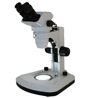
- First sortings are often performed with a stereo microscope.
- Inverted microscopes are used to assess morphology and since the observing objective needs to be close to them the inverted microscope does the trick.
- ICSI and IMSI (see below) require high resolution and high contrast microscopy.
- There are several types of reproductive medicine that microscopes are used for including:
In Vitro Fertilization (IVF)
- - This is a process where eggs are harvested and incubated, unselected sperm cells are added and left together (in vitro) for several days. Healthy mobile sperm cells actively fertilize the eggs and then embryos are transferred back into the uterus.
Intracytoplasmic Sperm Injection (ICSI)
- - This is a process where eggs are harvested and incubated, the oocyte is stabilized by a holding pipette, and a glass micropipette is used to collect a single sperm. The unselected sperm cell is immobilized by cutting its tail with the point of the micropipette. The oocyte is pierced through the membrane (oolemma) and the sperm is directed to the innter part of the oocyte (cytoplasm). The sperm is then released into the oocyte.
Intracytoplasmic Morphologically Selected Sperm Injection (IMSI)
- - This is a process where eggs are harvested and incubated. An inverted microscope is used for morphological selection of a healthy egg. The oocyte is stabilized by a holding pipette and semen is analyzed with an upright microscope. Morphological selection of a healthy sperm cell is performed with an inverted microscope with high magnifications, DIC and oil immersion objectives. A glass micropipette is used to immobilize the selected sperm by cutting its tail. The oocyte is pierced through the membrane (oolemma) and the sperm is directed to the inner part of the oocyte (cytoplasm). The sperm is then released into the oocyte. Cellular structures such as the zona pellucida and polar body of the egg cell must be clearly visible and the shape and vacuole count of the sperm cells must be assessed.
For details regarding any of our featured microscopes, contact us. You can also request a quote today.

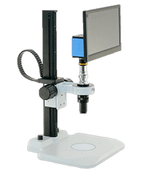
No eyepieces required
Visual Inspection Systems
These systems are perfect for quality control areas where a number of parts need examination throughout the day.
Shop Now
Same day response
Request a Quote
We build custom solutions tailored to fit your microscope needs. We respond to quote requests the same business day.
Submit Request


