Gout Microscopes
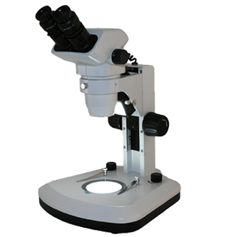
Medical professionals use gout microscopes assembled specifically for identifying gout or CPPD (pseudo-gout) crystals suspended in synovial fluid. Gout is a kind of arthritis that occurs when uric acid builds up in blood and causes joint inflammation. Gout is diagnosed by taking synovial fluid from the infected joint in the process of arthrocentesis. Lab technicians prepare a wet smear on a microscope slide with the fluid and use polarized microscopy to determine the presence of sodium urate crystals (gout) or calcium pyrophosphate dehydrate or CPPD within the fluid. CPPD crystals are small rods, squares, or rhomboids and are usually harder to identify without a gout or polarized light microscope. Polarizing filters can be easily adjusted when using the gout microscope. For the highest quality optics, the Zeiss gout microscopes will provide the best images. If a value brand is desired, the RB40 gout microscope provides optimal price-performance. These microscopes are used for identification of crystals present in body fluids. Contact Microscope World with any questions you have about a specific gout microscope setup.

No eyepieces required
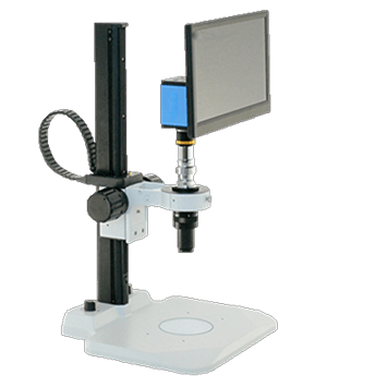
Visual Inspection Systems
These systems are perfect for quality control areas where a number of parts need examination throughout the day.
Shop Now
Same day response
Request a Quote
We build custom solutions tailored to fit your microscope needs. We respond to quote requests the same business day.
Submit Request


