Different Types of Microscopes Explained
The microscopes described below each create their image in a different way. The most commonly used microscope is the light microscope, which uses the light we can see to pass through a sample and produce an image.
5 Microscopes Create Images Using Different Techniques:
- Light Microscopes
- Confocal Microscopes
- Scanning Electron Microscopes (SEM)
- Helium Ion Microscopes
- X-Ray Microscopes
 Light Microscopes
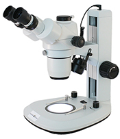
Light microscopes create the image you see using light. These microscopes are used in homes, schools, hospital labs, and in manufacturing facilities. The magnification of a light microscope is limited by the optical properties of light. Light microscopes include upright compound microscopes for studying a fixed sample, inverted microscopes for viewing cell cultures or live cells, stereo microscopes to produce a 3D image and view small parts, and zoom microscopes that provide higher resolution thanks to a higher numerical aperture in the lens.Confocal Microscopes
 Unlike the light microscopes mentioned above, a confocal microscope uses laser light as a light source. The laser scans the sample using different patterns, and the image is assembled with the computer. The laser can penetrate the sample deeper than light from a bulb. The resulting image is a three-dimensional image of controlled depth of field. A confocal microscope allows you to examine interior structures of cells, model organisms and tissue by stacking several images from different optical planes. Confocal microscopes can also be used for surface analysis in quality assurance and control.
 Scanning Electron Microscopes (SEM)
Electron microscopes use electrons rather than visible light to create an image. In order to use electrons for microscopy, you must focus the electron beam, and you need a vacuum. The beam is sent onto the sample, bounces off and creates a three-dimensional image of the surface. The wavelength of the electrons is much smaller than the wavelength of light from a bulb or a laser, resulting in higher resolution. When you want to use a scanning electron microscope, your sample must be electrically conductive, so the electrons bounce off of it. Samples used with scanning electron microscopes are often coated in a thin layer of gold or other metal.
The image below is a scanning electron image showing SARS-CoV-2 (in yellow), the virus that causes COVID-19. You can view more Covid images under the microscope here.

Photo credit: NIAID-RML.
Helium Ion Microscopes
 Helium ion microscopes are similar to a scanning electron microscopes in that the imaging technology is based on a scanning helium ion beam. The microscope allows the combination of milling and cutting of samples with their observation at sub-nanometer resolution. In terms of imaging, the helium ion microscope has several advantages over the scanning electron microscope. Due to the very high source brightness and the short wavelength of helium ions it is possible to obtain qualitative data not achievable with conventional microscopes that use photons or electrons as the emitting source. As the helium ion beam interacts with the sample, it does not suffer from a large excitation volume and therefore provides sharp images with a large depth of field on a wide range of materials. Helium ion microscope images provide information-rich images that offer topographic, material, crystallographic, and electrical properties of the sample with very little sample damage.
 X-Ray Microscopes
X-ray microscopes use electromagnetic radiation (x-rays) to produce images of tiny objects. Unlike a scanning electron microscope, the x-ray microscope can be used to generate an image of living cells. X-rays penetrate easier than visible light into the depth of a sample. X-ray microscopes can mirror the inside of samples non-destructively and create a three-dimensional image of the inner structure of an object with higher spatial resolution.
If you would like a quote for any of the above mentioned microscopes, please contact Microscope World.
